OCD Brain vs. Normal Brain:
Unveiling the Neurological Differences
The comparison between an OCD brain and a normal brain offers profound insights into the neurological underpinnings of Obsessive-Compulsive Disorder (OCD). While both types of brains share many common features, research studies have revealed distinct differences that shed light on the origins and manifestations of this challenging mental health condition. In this article, we will delve into these detailed differences, incorporating graphics and referencing relevant research studies, to elucidate the distinctions between an OCD brain and a normal brain.

Hyperactivity in the Orbitofrontal Cortex (OFC)
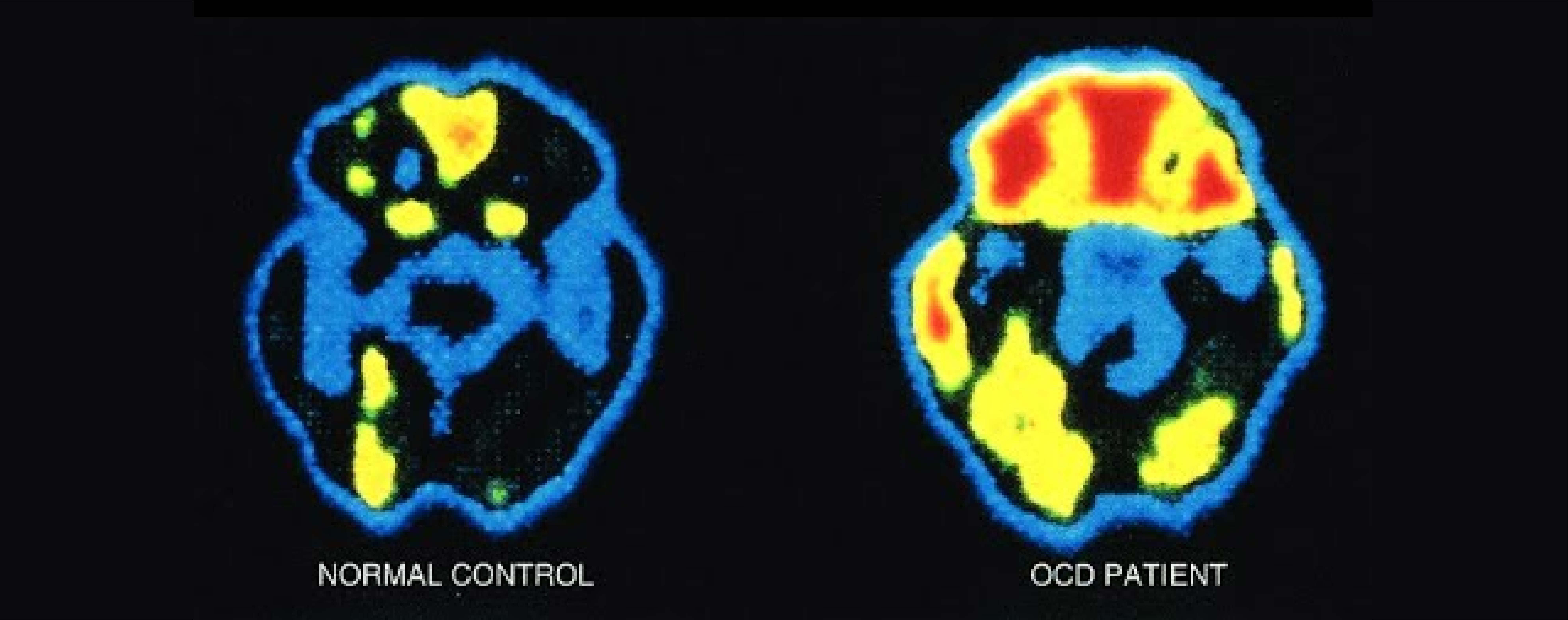
OCD Brain: Studies using functional magnetic resonance imaging (fMRI) have consistently shown heightened activity in the orbitofrontal cortex, a region involved in decision-making and impulse control. This hyperactivity is often associated with obsessive thought patterns in individuals with OCD.
Normal Brain: In contrast, a normal brain typically exhibits balanced activity in the OFC, without the excessive neural firing seen in OCD.
Altered Striatal Function
OCD Brain: The striatum, a brain region responsible for regulating habit formation and reward processing, shows
abnormal connectivity and function in individuals with OCD. Excessive activity in this area contributes to the compulsive behaviors characteristic of OCD.
Normal Brain: In a normal brain, the striatal function remains within the bounds of healthy habit formation and reward processing.
Dysregulated Serotonin Levels
OCD Brain: Research has revealed that individuals with OCD often have dysregulated serotonin levels. Serotonin is a neurotransmitter that plays a key role in mood regulation and anxiety management. The imbalance of serotonin can contribute to the anxiety and obsessive thought patterns in OCD.
Normal Brain: A normal brain maintains stable serotonin levels, helping to regulate mood and reduce anxiety.
Overactive Anterior Cingulate Cortex (ACC)
OCD Brain: The anterior cingulate cortex, responsible for error detection and emotional regulation, is overactive in individuals with OCD. This overactivity can lead to heightened self-criticism and anxiety.
Normal Brain: In a normal brain, the ACC functions within healthy parameters, enabling error detection and emotional regulation without excess.
Differences in White Matter Connectivity
OCD Brain: Recent studies using diffusion tensor imaging (DTI) have identified alterations in white matter connectivity patterns in OCD brains. These differences may contribute to impaired communication between brain regions responsible for decision-making and habit formation.
Normal Brain: In a normal brain, white matter connectivity remains more consistent and optimized for efficient
communication between regions.
Graphics and Visual Representation
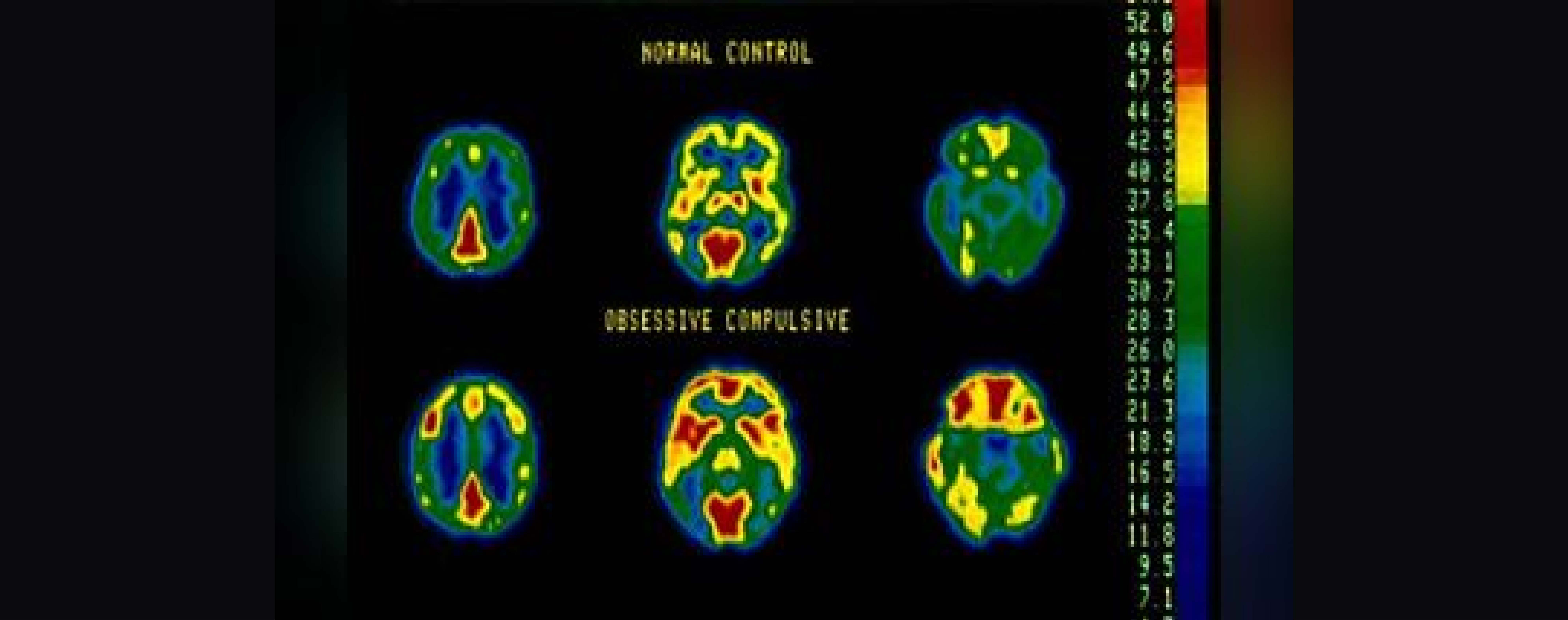
To visualize these differences, consider the following graphic from Yale school of medicine:
“The top row of brains here is from an individual without any diagnosis; the bottom row is from a patient with OCD. The images are reconstructed horizontal slices through the brain; the front of the brain is at the top, as if the person was lying on his or her back. The colors correspond to brain activity (if we allow a few technical assumptions that need not trouble us here). The warmer colors - reds and yellows - correspond to brain regions with higher activity; the cooler ones - blues and greens - are brain areas with less activity. By comparing the images in the top row with those in the bottom, you can see at a glance that, while the overall patterns are quite similar, a few brain regions markedly more active in the OCD brain. The two most prominent are the orbitofrontal cortex (OFC), at the front of the brain just above the eyeballs, and the caudate nucleus , a component of the basal ganglia deep within the brain.”

Conclusion
The comparison between an OCD brain and a normal brain highlights the intricate neurobiological differences that contribute to the development and persistence of Obsessive-Compulsive Disorder. Researchers continue to explore these distinctions to refine treatment approaches and develop targeted interventions for individuals with OCD.
Understanding these neurological differences underscores the complexity of OCD and the importance of a multidimensional approach to its treatment, including psychotherapy, medication, and neurobiological interventions. By exploring these differences, we move closer to unraveling the mysteries of OCD and improving the lives of those affected by this challenging condition.